
The program is now available
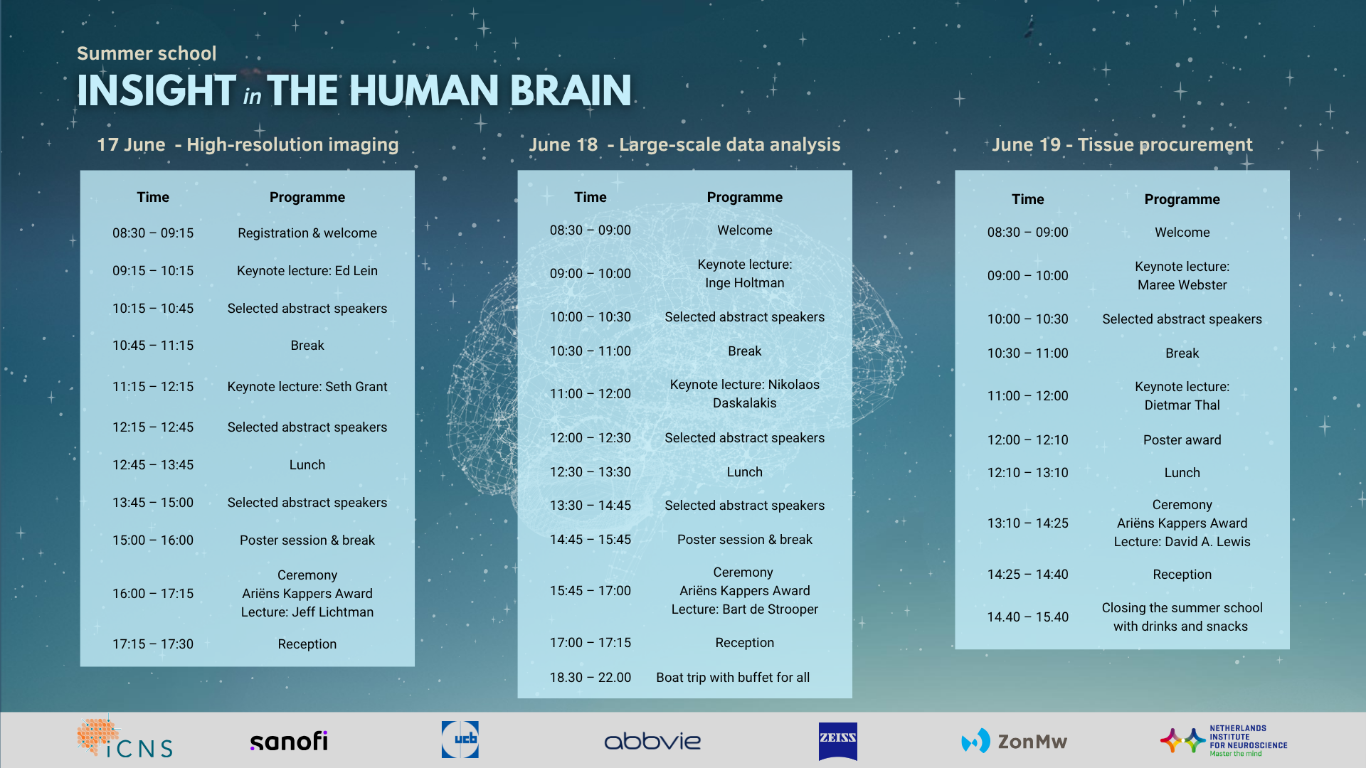
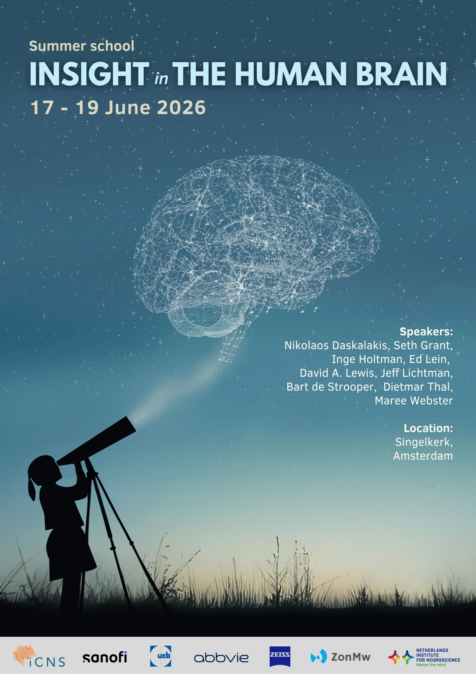
Every two years, the Netherlands Institute for Neuroscience (NIN) organises a Summer School that brings together leading researchers and early-career scientists in the field of neuroscience. This year’s edition, entitled “Insight in the Human Brain,” will take place from 17–19 June 2026 at the Singelkerk in Amsterdam.
Although the human brain has been studied for centuries, our ability to investigate its cellular and molecular organisation in human tissue has advanced dramatically only in recent years. Driven by breakthroughs in high-resolution imaging, spatial and single-cell omics, large-scale data integration, and AI-based analysis, human brain tissue research is rapidly transforming our understanding of brain function and disease. By organising the Insight in the Human Brain summer school, we aim to educate and update researchers on state-of-the-art experimental and computational approaches, while highlighting the importance of high-quality tissue procurement and detailed donor phenotyping. By bringing together leading international experts in neuroscience, neuropathology, imaging, and data science, the NIN summer school will provide a unique platform for training, networking, and in-depth exchange among researchers, students, and clinicians in this fast-evolving field.
Speaker Information
Bart de Strooper
Alzheimer’s Disease: connecting pathogenic mechanisms to hallmark symptoms.
The amyloid hypothesis, which posits that amyloid-beta accumulation drives Alzheimer’s Disease, has dominated the field for decades. However, mounting molecular, cellular, and therapeutic evidence challenges this linear, neurocentric view of disease progression.
Building on the concept of the “cellular phase of Alzheimer’s Disease,” we propose a model that highlights the central role of progressive gliosis and inflammation. These processes, initially triggered by amyloid fibrils, gradually become self-sustaining, driving neuropathology through distinct inflection points that mark successive phases of disease.
This theory is supported by the avalanche of novel insights generated over the past decade from transcriptomic and proteomic studies of sporadic AD cohorts. These findings emphasize the complexity of disease mechanisms, which unfold in parallel and necessitate phase-specific, tailored therapeutic strategies. The proposed model provides a unifying framework to integrate and interpret the wealth of data that has accumulated over the last decade.
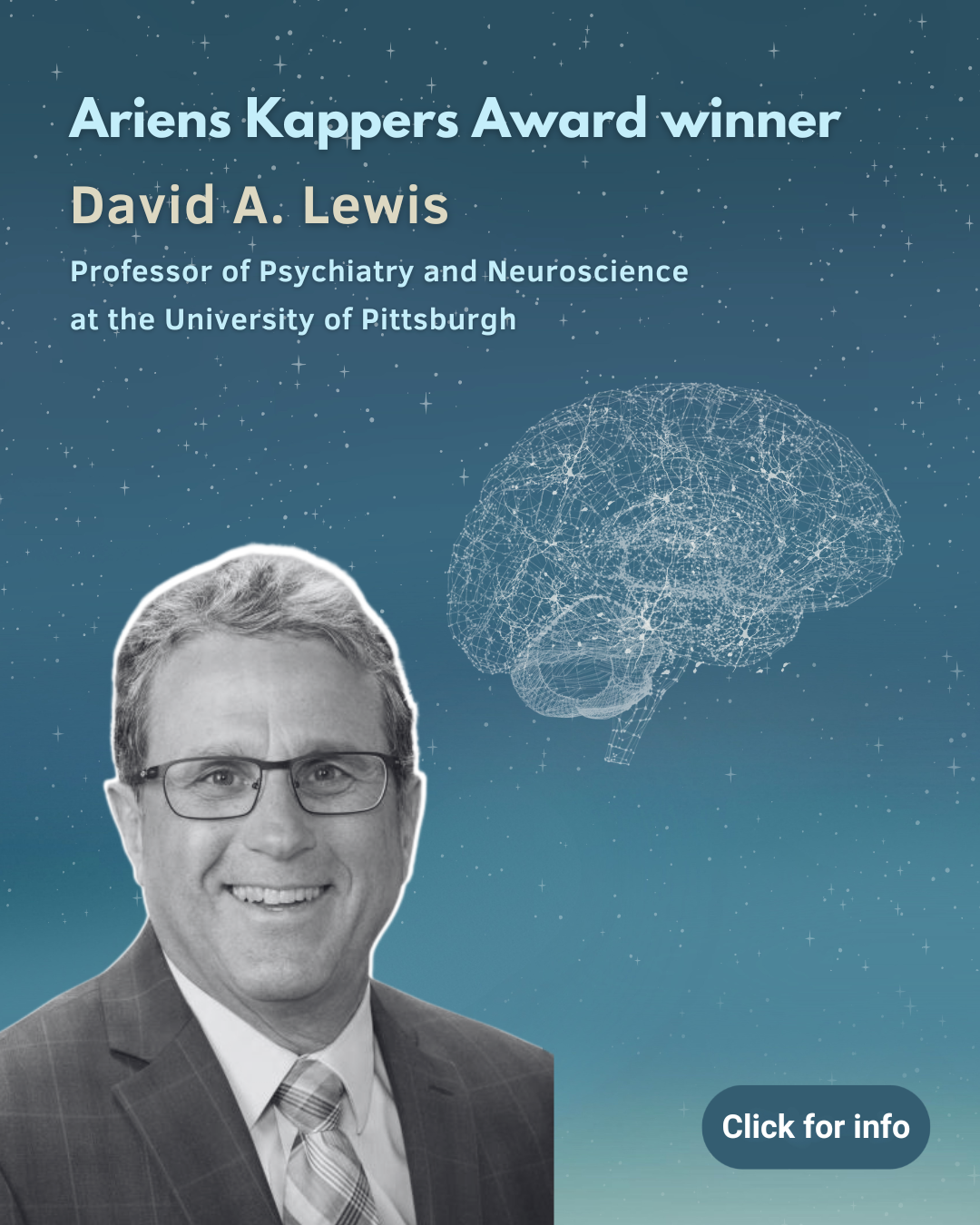
David Lewis

Cognitive deficits and cortical circuitry alterations in schizophrenia: The essential role of postmortem human brain studies
The locus of the disease process in psychiatric disorders, the human brain, is an appropriately privileged organ that is currently not subjected to biopsy in the diagnostic process of psychiatric illnesses for both clinical and ethical reasons. As a result, research in psychiatry has lacked the means to directly study the molecular and structural alterations of the brain at cell type-specific resolution in the living state, a key strategy that has led to major advances in understanding diseases of other organs that are biopsied. Consequently, studies of the postmortem human brain have received increasing attention as an essential vehicle for advancing our knowledge of the neurobiology of psychiatric illness. Using schizophrenia as an example, this presentation will illustrate strategies for combining studies of the postmortem human brain and of experimental model systems to 1) identify molecular, cellular and circuitry alterations associated with the core cognitive impairments of schizophrenia which currently lack effective treatments, and 2) determine if a given alteration in cortical circuitry likely represents a cause, consequence, or compensation in the disease process of schizophrenia…or a cooccurring factor (e.g., medications, substance use) or an experimental confound. The fundamental premise of these strategies is that by discovering the core pathological alterations in the diseased brain, we gain insight into both upstream pathogenic processes that arise from the convergence of a complex array of genetic and environmental disease risk factors and downstream pathophysiological processes which give rise to the emergent clinical features, such as cognitive deficits, that are characteristic of the diagnosis.
Dietmar Thal
Phenotyping quality of brain tissue and its relevance for research: Alzheimer’s disease as example.
Neuropathological phenotyping of brain tissue in brain banks and brain tissue resources delivers the backbone for any further analysis of the respective cases. Accordingly, the quality of this phenotyping will determine the quality of all further experiments that are done with the respective samples. When requesting samples from Alzheimer’s disease (AD) brain from a brain bank, you will receive (1) clinical information about the degree of dementia and additional diseases and (2) neuropathological information about amyloid and tau pathology (amyloid phases, Braak NFT-stage, and CERAD score for neuritic plaques). However, vascular pathologies, TDP-43, and α-synuclein (αSyn) lesions frequently occur in elderly brains with AD as co-pathologies. All these pathologies have impact on severity and subtypes of Alzheimer’s disease. For example, TDP-43 inclusions in neurons can colocalize with neurofibrillary tangles (NFTs). By doing so, abnormal tau can physically interact with phosphorylated TDP-43 (pTDP-43). This happens mainly in the subiculum and in the CA1 sector of the hippocampus. The tau levels in these cases increase in the brain and are associated with a pronounced loss of neurons in the subiculum and the CA1 sector by an increased activation of the necroptosis pathway in these cases. Another example is αSyn-positive Lewy body pathology of the amygdala-predominant type. This form of Lewy body pathology often lacks Parkinson-like Lewy body distribution patterns in the brain and contributes to the cognitive problems that AD patients develop. αSyn can occur in the same inclusions in neurons of the amygdala that also contain abnormal tau and pTDP-43. If this is the case, amygdala-predominant αSyn pathology is associated with even stronger neuron loss in the subiculum and the CA1 region. Finally, proteome changes can be linked to the presence/absence of TDP-43 pathology in AD cases.
Moreover, pathology distribution can vary among different brain regions. Of specific importance is here, to analyze best H&E-stained section of the regions requested for distinct tests to exclude the presence of infarcts and to determine the local pathology pattern. When using frozen samples for biochemical tests for which histological sections are not available, at least photo documentation of the macroscopic brain slabs is needed to exclude infarcts and bleeding. Pathology levels will need to be estimated in these cases according to the stages of pathology distribution.
Based on these examples, it becomes clear that the neuropathological phenotyping of biobanked samples determines which results can be found or not. Therefore, it is essential to make sure that the biobank can provide all clinical and neuropathological information that is required for a planned project before requesting the samples.
Ed Lein

From brain atlases to potential therapies: Deep cellular and molecular brain mapping, disease modeling and cell- and circuit-selective therapeutics
Remarkable advances in scalable cellular and molecular technologies to study the human brain are rapidly leading to the creation of comprehensive, multimodal, multiscale and cross-species foundational cell atlases through the NIH BRAIN Initiative Cell Atlas Network. Applied to Alzheimer’s disease these technologies and knowledge base now allow a detailed understanding of vulnerable and affected cell types along the course of disease. For example, specific subsets of cortical Somatostatin-positive GABAergic interneurons are the earliest vulnerable neuron types in Alzheimer’s disease, illustrating circuit-selective effects even at very low levels of pathology. Finally, these cell atlases allow the identification of short regulatory enhancer elements sufficient to restrict gene expression to specific cell populations with AAV-based delivery, opening new avenues for cell- and circuit-selective therapeutics which we demonstrate for genetic childhood epilepsies.
Inge Holtman
Decoding Brain Disorders: Integrating Genetics, Clinical Data, and Neuropathology in the Netherlands Neurogenomics Database
Neurodegenerative and psychiatric disorders share molecular mechanisms and genetic risk factors, often leading to clinical misdiagnoses. To achieve a data-driven understanding of the heterogeneity underlying these disorders, we established the Netherlands Neurogenomics Database, integrating clinical, neuropathological, and genetic data from over 3,000 brain donors collected by the Netherlands Brain Bank. Using Large Language Models (LLMs), we converted medical record summaries into longitudinal clinical trajectories that captured both known and novel disease-specific symptoms, enabling disease prediction and subtyping. In parallel, analyses of genome-wide variant data and polygenic risk scores (PRS) refined existing GWAS findings and highlighted shared as well as distinct genetic architectures across disorders. Together, these integrative approaches reveal converging and diverging biological mechanisms across brain disorders and pave the way toward more precise and personalized diagnostics.
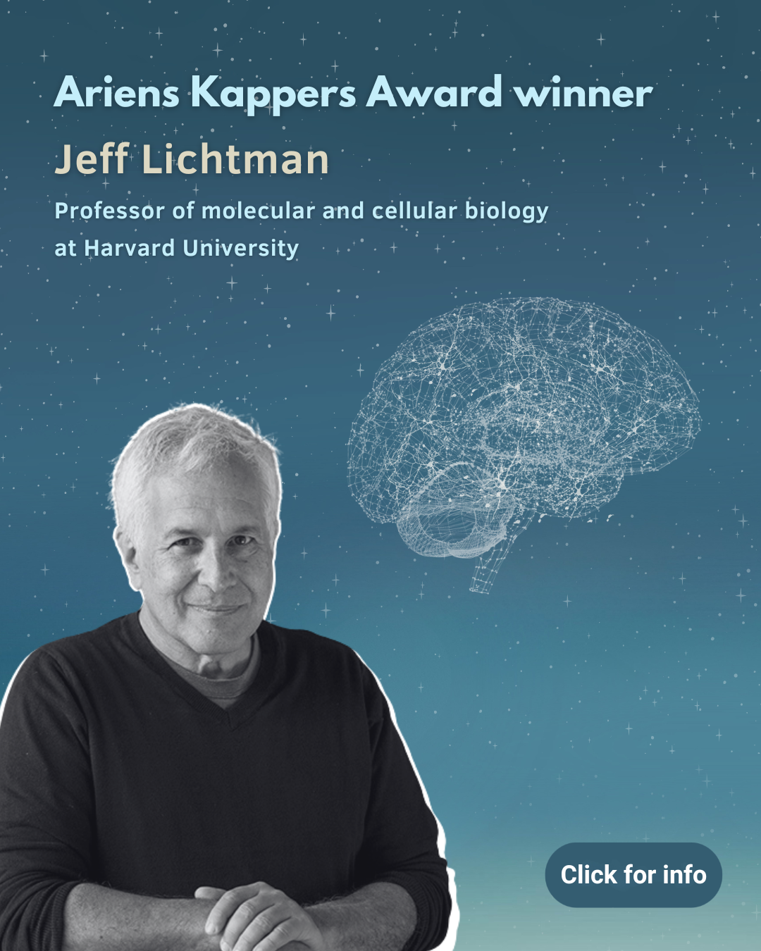
Jeff Lichtman

High-resolution Imaging of the Connectome
The nervous system functions through the action of an enormous number of synaptic connections between neurons. Making sense of this bewildering array of connections is the central challenge of connectomics, a synapse-level description of the brain’s wiring diagram. Using techniques that reveal this complexity, my lab colleagues and I have sought regularities in these connections to better apprehend the principles that underlie neural circuits. Comparisons of developing and adult circuitry in a range of vertebrate and invertebrate animals suggest that in animals, where learning is essential, structural circuitry motifs are products of experience, whereas in animals whose behaviors are more innate, comparable structural motifs are products of heredity.
Maree Webster
Maximizing the Utility of a Brain Bank
The Stanley Medical Research Institute (SMRI) Brain Collection was established to promote and facilitate research on schizophrenia, bipolar disorder and major depression. The samples are distributed free of charge to qualified researchers. All studies are conducted on the same specified cohorts of demographically matched cases and controls. The samples are distributed coded so that researchers must return all data to SMRI before they can obtain the demographic and clinical information for each subject. By distributing slide based, fixed and frozen sections and aliquots of RNA, DNA, and protein, thousands of studies can be conducted on each brain. Anatomical maps of heterogenous regions such as the thalamus are made to enable accurate dissections of specific subregions and to ensure anatomical matching of samples across cases. To date, SMRI has acquired over 7,000 individual data sets, as well as numerous high throughput, genomic, transcriptomic and proteomic data sets from multiple brain regions and from peripheral samples from each case. All data is available through the SMRI website and can be used to validate or corroborate findings and to generate additional hypothesis.
Nikos Daskalakis

Systems Biology of Stress related mental disorders
The Daskalakis Neurogenomics and Translational Bioinformatics Laboratory (NG-TBL) investigates the molecular, cellular, and genomic pathways linking stress to brain function and dysfunction. NG-TBL uses functional genomics to dissect epigenetic mechanisms, cell-type-specific regulatory elements, and risk loci that contribute to stress-related mental disorders such as PTSD. Translational studies using cell-based and animal models further validate causal genes and variants emerging from human genomics. The laboratory also employs large-scale datasets and machine-learning approaches to map genotype-to-phenotype relationships in complex neuropsychiatric traits, with the goal of understanding individual differences in symptom expression and vulnerability. NG-TBL’s overarching mission is to identify novel mechanisms and therapeutic targets for stress-related mental disorders.
Seth Grant

The architecture of synapse diversity in health and disease
The brain’s defining feature is its enormous number of synaptic connections. Traditionally, synapses were thought to fall into only a few broad classes—excitatory, inhibitory, or modulatory—based largely on neurotransmitter identity. The advent of synaptome mapping, which enables systematic, single-synapse–resolution analysis across the entire mouse brain, has overturned this view. These studies reveal an unexpectedly vast diversity of synapse types and their organisation into a highly structured three-dimensional map known as the synaptome architecture. This architecture provides a powerful new framework for understanding how innate and learned behaviours are encoded, how neurological and psychiatric diseases selectively target specific synapse populations, and how therapeutic drugs act on brain circuits.
More Information
Who is the summer school open to?
Advanced students and professionals in neuroscience and related disciplines, including neuroscientists, biologists, psychologists, and clinicians. All participants are expected to have a minimum of bachelor-level knowledge in neuroscience, psychology, or a related field.
What are the participation costs?
The registration fee is €450, including lunch, reception & boat tour. We aim to accommodate approximately 170 attendees.
Who are the confirmed speakers?
Nikolaos Daskalakis, Seth Grant, Inge Holtman, Ed Lein, David A. Lewis, Jeff Lichtman, Bart de Strooper, Dietmar Thal, Maree Webster.
What topics does the programme cover?
The programme will feature lectures by internationally renowned scientists and award recipients at the forefront of human brain research. They will present their work across three main themes: the analysis of human brain tissue, the utilisation of large data sets, and the procurement of human brain tissue for research. In addition oral presentation will be selected from the abstracts.
Who are the members of the organising committee?
Netherlands Institute for Neuroscience: Inge Huitinga, Maarten Kole, Evgenia Salta, Chris de Zeeuw
Where does the event take place?
Singel 452, 1017 AW, Amsterdam, the Netherlands
How can I get more information?
For further details, please contact the organisers by e-mail at: summerschool2026@nin.knaw.nl
Ariens Kappers Award Winners

About AbbVie
![]()
AbbVie’s mission is to discover and deliver innovative medicines and solutions that address serious health issues today and the medical challenges of tomorrow. We strive to have a remarkable impact on people’s lives across several key therapeutic areas including immunology, neuroscience and oncology – and products and services in our Allergan Aesthetics portfolio. In Germany, AbbVie is represented by its corporate headquarters in Wiesbaden and its research and manufacturing site in Ludwigshafen. Globally, AbbVie employs approximately 57,000 people, including around 3,500 in Germany. For more information about AbbVie, please visit www.abbvie.de or follow us on LinkedIn.
Support our work!
The Friends Foundation facilitates groundbreaking brain research. You can help us with that.
Support our work