
At the imaging core facility of the NIN, we offer a wide array of light microscopy imaging modalities to support the basic, and advanced imaging requirements of neuroscience. With these systems researchers at the NIN are able to capture (sub)cellular structures and processes in the healthy and diseased brain in various ways.
We provide in-depth training for the usage of the microscopes, and also advice on tissue preparation, experimental design, and image analysis.
If you would like to use our equipment as an external researcher, feel free to get in touch!
Overview of equipment:
- Abberior STEDYCON super resolution (STED) microscopy attached to a Zeiss axiovert 200M microscope. For sub-diffraction-limit resolution imaging. Controlled with stedycon software.
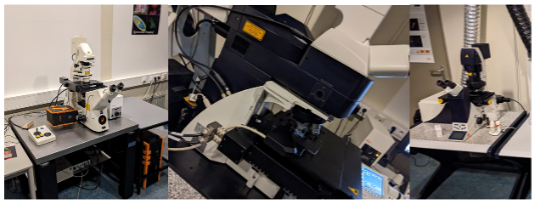
- SWCs’ BrainSaw serial two-photon tomography (STPT) microscope, for imaging whole mount samples.
- Leica TCS SP8 X confocal system on an upright DM6000, with 405 nm excitation laser and a white light laser (470-670nm), 1 PMT and 2 HyD detectors.
- Leica TCS SP5 II confocal system on an inverted DMi6000 research microscope with 405, 458, 488, 496, 514, 561, 594, and 633 nm Exc. lasers.
- Zeiss (Axio Scan Z1) slide scanner, 5x, 10x, 20x (0.8 & 0.45 NA), 40x objectives. Orca Flash 4.0 & Hitachi HV-F203SCL cameras, and colibri 7 illumination. For high-throughput imaging of slides with brightfield, and fluorescence.
- Zeiss Laser Microdissection microscope (Palm Microbeam) with 354 nm laser for micro dissection and collection of single cells or small regions from tissue, equipped with fluorescence (Exc. 400, 490, 590 nm). PALMRobo (Zeiss) software.
- Zeiss Axioskop brightfield upright microscope with scanning stage, and Qimaging Micropublisher 5.0 color camera. For high resolution transmitted light imaging applications. With imageProPlus 6.3 software.
- Zeiss Axiovert 200m inverted widefield fluorescence microscope, with scanning stage Qimaging camera. Fluorescence filters for excitation with 365, 480, 550 and 647 nm.
- Keyence VHX-X1 digital microscope for 2D/3D imaging and analysis of materials and electronics.
- Flatbed scanner Epson perfection v700 for larger specimens.
- Multiple high-performance workstations for data analysis with opensource software (Qupath, Fiji, Napari) and commercial software (Neurolucida).
Contact:
Support our work!
The Friends Foundation facilitates groundbreaking brain research. You can help us with that.
Support our work