Neurons can look outside the box
28 August 2023

28 August 2023
A new study from the Netherlands Institute for Neuroscience shows that individual neurons that do not receive meaningful visual input are still aware of the context through information communicated by other cells.
The efficiency of our vision is a remarkable achievement of evolution. The introspective ease with which we perceive our visual surroundings masks the sophisticated machinery in our brain that supports visual perception. When we process visual information, all cells in the visual cortex focus on a small snippet of the overall image. It’s as if you’re presented with a palette of cells that initially appear to deal with tiny fragments. There isn’t a single cell where this information converges.
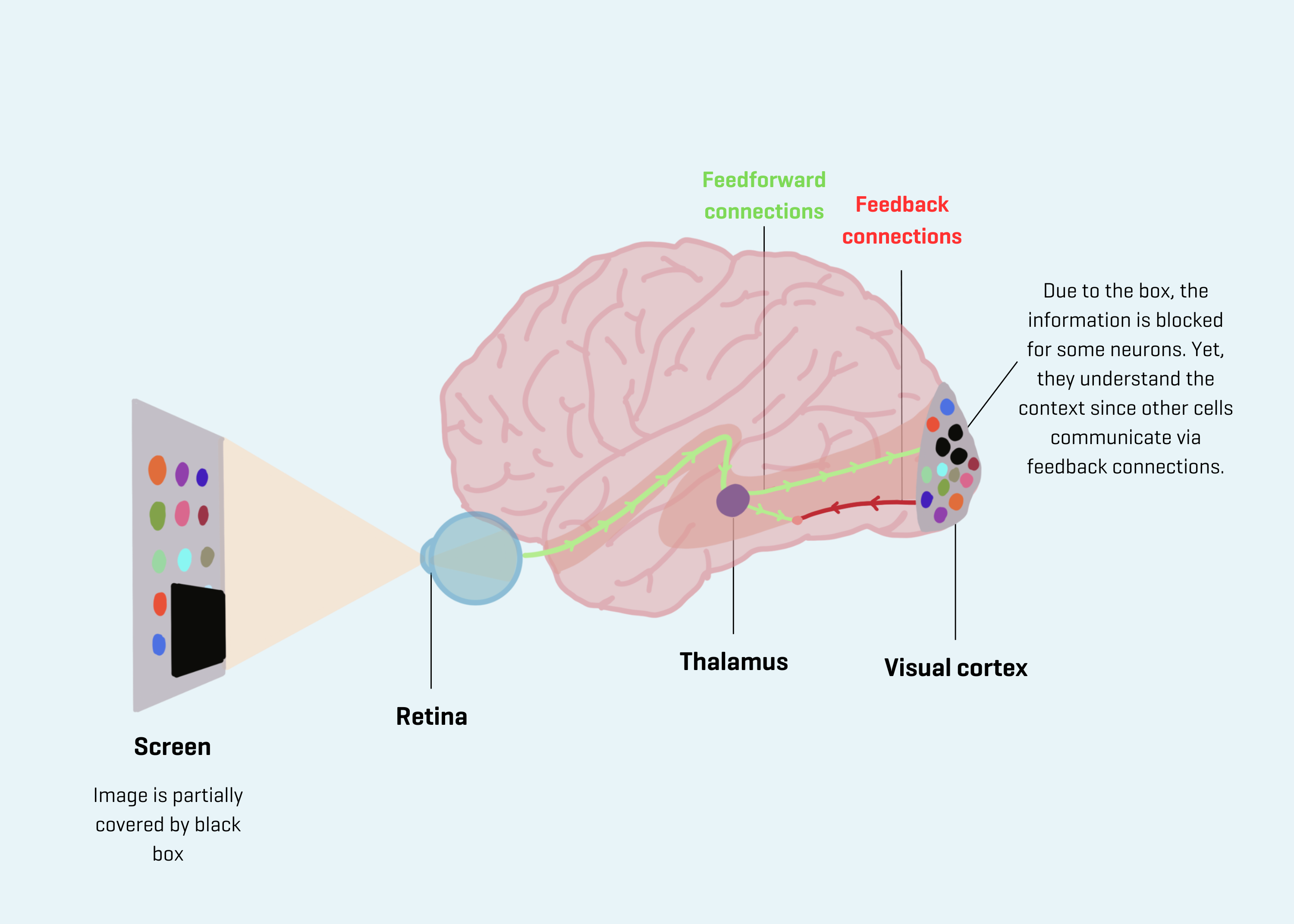
Visual input travels through the retina to the thalamus (the relay station for sensory impulses), and is then transferred to the visual cortex. The specifics of what neurons in this area see are regulated by these so-called ‘feedforward connections’ or direct visual input. In addition, neurons also receive ‘feedback connections’ that provide more information about the context of the image. But what happens exactly and how does this work?

Figure visual system: Screen showing an image that is partly hidden by a black box. The retina receives visual input and sends this information to the thalamus, the relay station for sensory impulses. This information is then transferred to the visual cortex by feedforward connections. Each cell focuses on its “receptive field”, which is a small snippet of the overall image. Because of the box, the information is blocked for some neurons. However, it turns out that they still understand the context, due to feedback from higher brain regions.
Previous studies attempted to explore this in humans by showing images of visual scenes, which were partly covered by a black box. While for some cells in the visual cortex the location is covered, it seems that they somehow grasp what’s happening in the image and its context through other pathways. It’s as though they’re hearing this from other cells. To gain deeper insights into this, research at the cellular level is necessary. However, in humans, observing individual cells in the living brain is challenging.
For this reason, Paolo Papale, Feng Wang, and their colleagues, under the supervision of Pieter Roelfsema, conducted similar experiments in macaque monkeys. Paolo Papale says, “We have monkeys looking at a screen with various images. Some of these images were normal scenes (like a picture of a beach), and some had a black box covering part of the image. Next, we measured the activity of 175 neurons in the primary visual cortex. These cells are responsible for a small part of the visual field. By blocking part of the image, theoretically, some of the cells wouldn’t know what’s in the image. Surprisingly, however, they did seem to know. Our results suggest that these neurons were informed by feedback from higher visual brain regions where there are neurons that process information about the non-occluded parts of the scene.’
“In the future, we want to delve into what information these feedback pathways are processing exactly. In the case of an image of a forest, for instance, do they send information about how many trees are there? What do they transmit and what don’t they transmit? These results show that individual cells in the visual cortex don’t merely function as pixels; they also respond to context. This demonstrates that what we see isn’t objective. It doesn’t work like a camera with pixels: context matters. This can also relate to why different people have different perceptions, even when looking at the same thing.”
Source: Current Biology
Watch the video abstract here

The Friends Foundation facilitates groundbreaking brain research. You can help us with that.
Support our work