The white matter of the MS brain shows abnormalities even before inflammation
25 May 2023

25 May 2023

Patients with MS show structural abnormalities in their white matter even before MS inflammation develops. This is the conclusion of a new study by the Netherlands Institute for Neuroscience (NIN) in Amsterdam and the Max Planck Institute for Multidisciplinary Sciences in Göttingen (MPI). Could this finding be a target for a new treatment to prevent MS inflammation?
Multiple sclerosis (MS) is a chronic inflammatory disease of the central nervous system. In early, as well as advanced progressive MS, lesions arise along with substantial inflammatory activity. Lesions are the inflammatory sites where the myelin is broken down and taken up by microglial cells (our brain’s immune cells). But do we see something in the tissue even before these inflammation spots appear?
To answer this question, Aletta van den Bosch of the research team of Inge Huitinga (NIN) and Wiebke Moebius (MPI) looked into human post-mortem brains of MS patients and controls that have been donated to the Dutch Brain Bank. Their focus was particularly on the so-called ‘normal-appearing white matter’. As the name suggests, these are areas where lesions have not formed yet, and so still appear normal. How is it possible that people with MS develop lesions here later and people without MS do not?
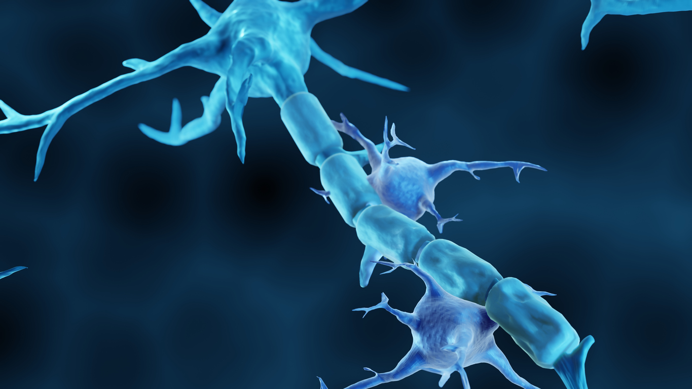
The team took a detailed look at myelin to see if there are early changes in people with MS. Myelin is an insulating white, fatty substance that is wrapped up to 150 times around the nerve fibers. At regular distances from each other there are interruptions of the myelin, these are the Nodes of Ranvier. During the transmission of electrical signals, the signal jumps from one Node or Ranvier to the next, allowing a myelin-containing fiber to transmit a signal 100 times faster than without myelin. In people with MS, myelin is damaged and signal transmission in the central nervous system is disrupted, which can impair functions such as walking and vision. What kinds of changes in brain tissue can we observe in the early stages of MS?
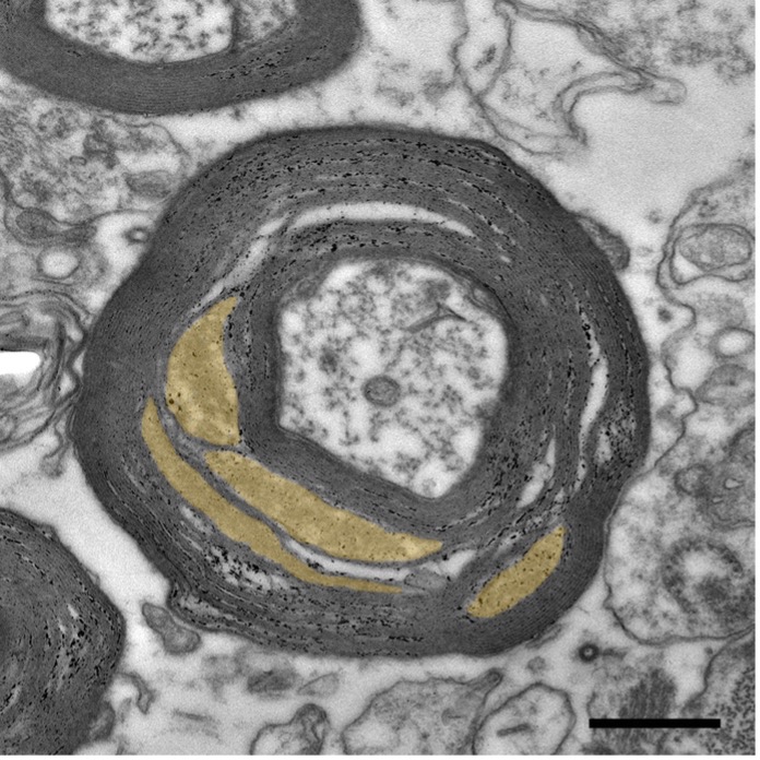
Aletta van den Bosch: ‘To be able to study myelin properly, we looked at the optic nerve. In this area, all nerve fibers and their myelin follow the same direction very nicely, so that we can visualize the myelin well. We did this by using electron microscopy. With this technique we zoomed in 5,000 to 30,000 times on a cross-section of a nerve-fiber.’
‘In MS, myelin was found to be less tightly wrapped around the nerve-fiber. This means that the fiber is not properly insulated which has major consequences: the signal can’t be transmitted as fast as it used to be. We saw that where myelin was less attached to the fiber, there was a disruption of the nodes of Ranvier combined with increased levels of T-cells and activated microglia. Furthermore, there were more mitochondria. Mitochondria are the energy factories of the cell, so this phenomenon may indicate that more energy is needed for signal movement and maintenance of the fibers.’

Figure: Electron microscope image of a nerve fiber with myelin (in black) in the optic nerve of an MS patient. In yellow, the spaces between myelin layers that are enlarged in MS) (Credit: W. Möbius, Max Planck Institute for Multidisciplinary Sciences)
‘Although mitochondria are generally good for energy production, they also produce many by-products, such as oxygen radicals. We suspect this to be an amplifying factor for myelin breakdown: the myelin is already in a bad state, more mitochondria develop to provide more energy, which then makes conditions even worse. The theory is that a threshold value is needed to initiate the breakdown. It is also possible that the body recognizes the detached myelin as ‘abnormal’, which could be the start of breakdown by immune cells.’
‘We haven’t been able to look at human tissue in such detail before, meaning that almost all the research so far has been done in laboratory animals. Although this is very valuable research, it could sometimes be more difficult to translate the results directly to humans. This is the first glimpse into what happens at the ultrastructural level in people with MS and what exactly leads to the lesions. You need very good tissue to do this which is why the brain bank is so crucial for our research.”
‘The next step is to see if we can prevent the myelin from winding so loosely around nerve endings. First, we want to experiment in culture dishes to see if we can make the wrapping of myelin stronger. Subsequently, we will have perform tests in laboratory animals, and eventually we will be able to take the step to humans. It would be great if we could find something to prevent myelin detachment. While this will not prevent the damage of the lesions that are already there, it might prevent the development of new lesions. This would provide a whole new target for MS treatment.”
Source: Annals of Neurology
Contact: Aletta van den Bosch (a.v.d.bosch@nin.knaw.nl) and Inge Huitinga (i.huitinga@nin.knaw.nl)

The Friends Foundation facilitates groundbreaking brain research. You can help us with that.
Support our work